Immunophenotyping Utilizing 6 Color Flow Cytometry
Introduction / Rational
Beginning on December 5, 2011, the Warde Medical laboratory’s flow cytometry section changed from a 4-to a 6-color analysis for evaluating for leukemias and lymphomas. This now allows for the full utilization of the new generation of flow cytometers acquired over the last several years.
The major advantages associated with this change are:
- fewer tubes, hence faster analytic throughput & greater labor efficiency,
- fewer duplicate (non-billable) antigens in a panel and
- more modern alignment with current recommendation for flow immunophenotyping of hematolymphoid neoplasias.
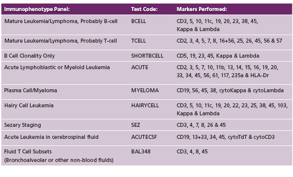
Table 1. Summary of Immunophenotyping Panels Available by 6-Color Flow Cytometry

CHANGES
1. Mature B Lineage Neoplasias
B Lymphoid
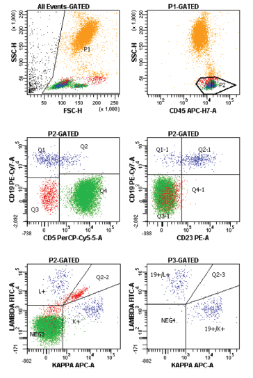
The current panel content for B cell clonality (Order code SHORTBCELL), Mature B cell lymphoma/leukemias (order code BCELL) and for Hairy Cell leukemia (order code HAIRYCELL) are unchanged in content. However, the number of tubes are essentially cut in half, with a significant reduction in duplicate antigens. The increased combination of simultaneously analyzed antigens allows for a more sensitive evaluation of minor B cell monoclones, as well as clearer identification of major lymphoid subsets (eg, T, B, NK, see Figure 1).
Plasma Cell Dyscrasias
Flow analysis for plasma cell dyscrasia is not routinely needed for diagnosis — morphologic exam of the marrow along with serum & urine protein electrophoresis / immunofixation are typically sufficient. However, flow analysis can be a particularly useful application in the setting of a known minor monoclonal gammopathy (< 3.0 g/dL) when there are relatively few plasma cells present in the morphologic specimen. The new plasma cell panel is designed to efficiently identify a small fraction of plasma cells and ascertain if they are monoclonal, polyclonal or a mixture of both — all in a single tube (order code MYELOMA).
2. Mature T Lymphoid Neoplasias
Mature T Cell Panel
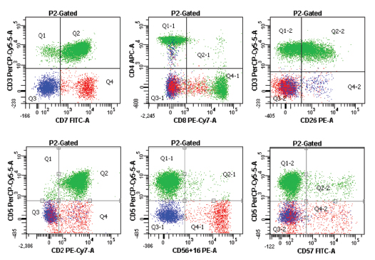
Similar to the B lymphoid panels, the mature T cell lymphoma/leukemia panel (order code TCELL) has reduced by half the number of tubes needed, while allowing for a greater ease of directly visualizing coexpressed T and NK associated antigens (see Figure 2). In order to accommodate a new T related antigen (CD26) and/or substitute in other T related antigens (eg, CD10 with a differential of angioimmunoblastic T NHL), the NK antigens CD16 and CD56 are combined. A follow up analysis separating these two antigens can be performed if desired.
Sezary Staging Panel
Tube #1 of the T cell panel can be used as a stand-alone staging analysis for involvement of the blood by a Cutaneous T cell lymphoma (ordercode SEZ). This evaluates for immunophenotypic abnormalities associated with these types of T cell neoplasias (eg, excess CD4+ T cells; loss of CD7 or CD26 on the CD4+ T cells). It is compliant with international staging protocols for cutaneous T cell lymphomas and Sezary syndrome.
3. Acute Leukemia & MDS
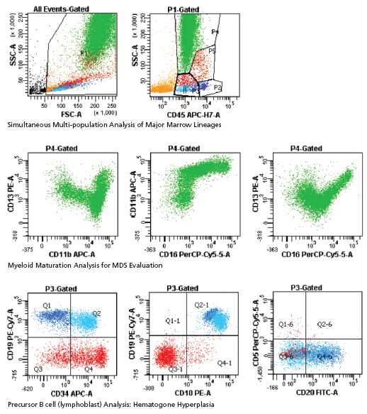
The new acute leukemia panel (order code ACUTE) is designed to more fully evaluate for acute leukemia as well as other causes of marrow failure and associated cytopenias (eg, myelodysplasia; B lymphoproliferative disorders). This includes a fuller ability to evaluate for visualization of atypical antigen expression on both mature B and T lymphs (see Figure 3). Lastly, if desired, an evaluation of cytoplasmic antigens to define major lineage (T vs. B vs. myeloid) is available as a single tube addition to the standard acute leukemia panel (cyto. TdT/cyto. MPO/ cyto. CD3/cyto. CD79a).
4. CSF Fluids
Flow immunophenotypic analysis of CSF for leukemia or lymphoma is really only indicated in patients with (1) a past medical / laboratory history of leukemia or lymphoma or (2) highly suggestive imaging or CSF cytologic evidence hematolymphoid neoplasia (eg, elevated or atypical lymphs; blasts).
In the usual clinical setting in North America, the major differential diagnoses will be between a mature B cell neoplasm and an acute leukemia. As such, there are two new panels — each a single tube — to evaluate for one or the other. The B cell clonality (order code SHORTBCSF) panel is the same as for non-CSF samples, and allows identification of major lymphoid subsets and B cell clonality and differentiation in one tube.
In the setting of suspected acute leukemia, a single tube panel designed to detect T & B lymphoblastic and AML is used (order code ACUTE CSF), using a mixture of membrane and cytoplasmic antigens. The specificity of the final diagnosis will be critically dependent upon correlation with other clinical and lab data.
Lastly, CSF samples reasonably submitted for flow analysis should be immediately processed and submitted in a 1:1 dilution of tissue media (eg, RPMI; McCoy’s).
Figure 1: B cell Analysis

Figure 2: T cell Analysis

Figure 3: Acute Leukemia/MDS panel


